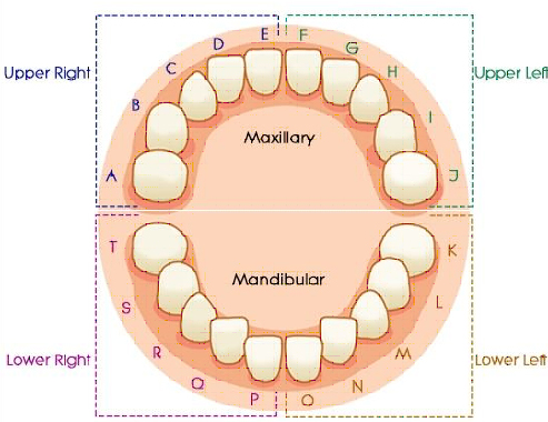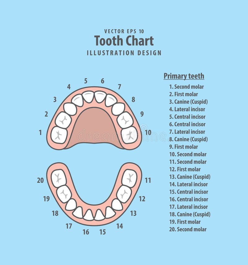

There are several notation systems used in dentistry to identify teeth. Roots are greatly reduced in size and can be fused together. The crown usually takes on a rounded rectangular shape that features four or five cusps with an irregular fissure pattern. Mandibular (lower) third molars are the smallest molar teeth in the permanent dentition. Their roots are commonly fused together and can be irregular in shape. Maxillary (upper) third molars commonly have a triangular crown with a deep central fossa from which multiple irregular fissures originate. Morphology of wisdom teeth can be variable. Structure ģD CT of an impacted wisdom tooth near the inferior alveolar nerve Tooth morphology

Some oppose the prophylactic removal of disease-free impacted wisdom teeth, including the National Institute for Health and Care Excellence in the UK. Yet, impacted wisdom teeth are commonly extracted to treat or prevent these problems. Some more conservative treatments, such as operculectomies, may be appropriate for some cases. Wisdom teeth which are partially erupted through the gum may also cause inflammation and infection in the surrounding gum tissues, termed pericoronitis. Impacted wisdom teeth may suffer from tooth decay if oral hygiene becomes more difficult. Impacted wisdom teeth are still sometimes removed for orthodontic treatment, believing that they move the other teeth and cause crowding, though this is not held anymore as true. Wisdom teeth may get stuck ( impacted) against other teeth if there is not enough space for them to come through normally. Most adults have four wisdom teeth, one in each of the four quadrants, but it is possible to have none, fewer, or more, in which case the extras are called supernumerary teeth. The age at which wisdom teeth come through ( erupt) is variable, but this generally occurs between late teens and early twenties. The teeth do not have any lymphatic vessels.A third molar, commonly called wisdom tooth, is one of the three molars per quadrant of the human dentition. Via vessels that generally follow the arteries. Venous drainage of the teeth is into either the: Inferior alveolar artery: arises from the maxillary artery then enters the mandibular foramen

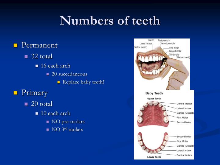
Middle superior alveolar artery: small branch of the infraorbital arteryĪnterior superior alveolar artery: branch of the infraorbital artery Posterior superior alveolar artery: branch of the maxillary artery in the pterygopalatine fossa Arterial supplyĪrterial supply to the teeth is derived from the maxillary artery, a branch of the external carotid artery, via the: This joint between a tooth and alveolar bone is a fibrous joint called a dentoalveolar syndesmosis. The periodontal ligament connects the tooth root to the underlying lamina dura, which itself is the cortical bone which lines the tooth socket. Pulp chamber and root canal: lie centrally within the tooth and contain neurovascular structuresĪpical foramen: lies at the apex of the tooth root Each tooth is mainly composed of dentin and is made up of several parts 1-3:Ĭrown: portion of the tooth projecting out of bone The tooth sits in alveolar processes of the upper jaw ( maxilla) or lower jaw ( mandible). The dental arch describes the crescentic formation of teeth on each jaw. There are normally a total of 32 permanent (secondary) teeth in adults, with 16 per jaw and eight in each quadrant, which consists of (distal to mesial) 3:

They are then progressively replaced by permanent (secondary) teeth from the age of six with the final eruption of the third molar between 18-24 years 5. The deciduous (primary) teeth start erupting at six months (lower central incisor) and are completely erupted by around 3 years of age. There are twenty deciduous (primary) teeth in young children, with ten per jaw and five in each quadrant, which consist of (distal to mesial):Ĭentral incisors are the first to erupt, around 6 months of age


 0 kommentar(er)
